
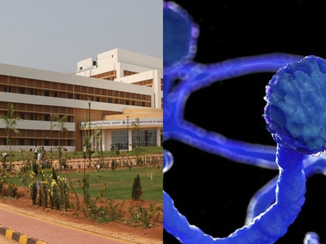
Bhubaneswar: AIIMS Bhubaneswar researchers has discovered a new fungus reportedly causing Mucormycosis and named it as Cunninghamella Arunalokei.
Cunninghamella arunalokei, a new species of Cunninghamella from India, can cause the disease in an immonocompetent individual.
Mucormycosis is an angioinvasive disease often associated with high morbidity and mortality. Infections due to Cunninghamella species are relatively rare in India in comparison to other countries.
Patients with hematological malignancy and hematopoietic stem cell transplant have an increased risk of acquiring mucormycosis due to Cunninghamella species which predominantly presents as pulmonary and disseminated infections. Cunninghamella species are associated with high mortality compared to Rhizopus species. A high mortality rate is possibly due to the pathogen’s virulence
compared to other Mucorales.
A 26-year-old male from Odisha came with erythema, induration, and superficial ulceration with crusting on the left dorsum of the nose and swelling in the left mid-half of the face. Histopathological examination report of the biopsied lesion and radiological imaging had confirmed the diagnosis of invasive mucormycosis with involvement of the facial skin, subcutaneous tissue, and the maxillary sinuses.
His left ala was destroyed, exposing the nasal septum. His laboratory investigations revealed normal hemogram, blood sugar levels, liver, and renal function parameters. Urine ketone bodies were not detected.
Serological examination for HIV was non-reactive, and the lymphocyte CD4 counts were in the normal range. His immunoglobulin and complement levels were also within normal limits. The lesions were biopsied again, and he was started on a combination of super saturated potassium iodide solution (SSKI) and oral itraconazole, considering a probability of atypical entomophthoramycosis. A potassium hydroxide mount of the biopsy showed broad aseptate hyphae, and culture grew a white cottony mold at 28 ◦C after 5 days of incubation. Lactophenol cotton blue examination of the mold demonstrated erect sporangiophores ending in a swollen vesicle with 1-spored sporangiola suggestive of Cunninghamella species.
A fresh biopsy confirmed a disease recurrence and the patient was treated with a combination of oral posaconazole and terbinafine for
6 months. The lesions regressed, and he was advised to continue oral terbinafine indefinitely. However, the patient could not procure terbinafine during COVID-19 pandemic lockdown for around 7 months and reported 2 months later to the emergency with disorientation, severe weakness due to reduced oral intake secondary to palatal fistula and nasal regurgitation.
A repeat biopsy culture performed confirmed recurrence. Magnetic resonance imaging revealed disease extension to anterior cranial fossa with skull base erosion The patient was initiated on liposomal amphotericin. A total of 4 g of liposomal amphotericin B was administered during four weeks. The patient improved gradually, and the lesion subsided. The patient was discharged on oral posaconazole 300 mg/day. However, the patient did not take the posaconazole due to financial constraints and succumbed to the illness 1 month after discharge.

 Ms Kalinga